
July's Case of the Month - 2023
Subcutaneous Foreign Body
Signalment:
Age: 8 year old
Gender: Neutered Male
Species: Canine
Breed: Doberman
History:
Abdominal ultrasound was requested for patient due to persistent fever, lethargy, anorexia and weight loss, and polydipsia. Previously performed thoracic and abdominal radiographs and bloodwork were all unremarkable.
With patient in dorsal recumbency for abdominal ultrasound, a subtle flat, firm, fluctuant swelling was noted over the right caudolateral ribs.
Ultrasonographic findings:
The subcutaneous tissue over the right caudal ribs is severely hyperechoic and surrounds pockets of echogenic fluid. There is a thin hyperechoic linear object (at least 4.4cm long) that extends from within a fluid pocket to between the 9th and 10th rib and penetrates into the abdominal cavity. Some free gas is seen within the fluid pocket.


Mesentery throughout the right cranial abdomen is diffusely hyperechoic.
The remainder of the full abdominal ultrasound was unremarkable.
Abdominal ultrasound interpretation:
Subcutaneous fluid pockets - the findings are moderate and consistent with abscess formation
Linear object - the findings are moderate and suggestive of a foreign body penetrating through the subcutaneous tissues and into the abdominal cavity
Mesentery - the findings are moderate - DDx: peritonitis - inflammation vs. paraneoplastic reaction vs. infectious vs. fibrosis vs. other.
Provided Recommendations:
An aspirate of a subcutaneous fluid pocket was recommended and performed. Purulent fluid was obtained confirming the presence of an abscess
Consider submitting aspirate for culture and sensitivity
Recommend exploratory laparotomy to fully evaluate the abdomen and foreign body and potentially resolve with surgical intervention.
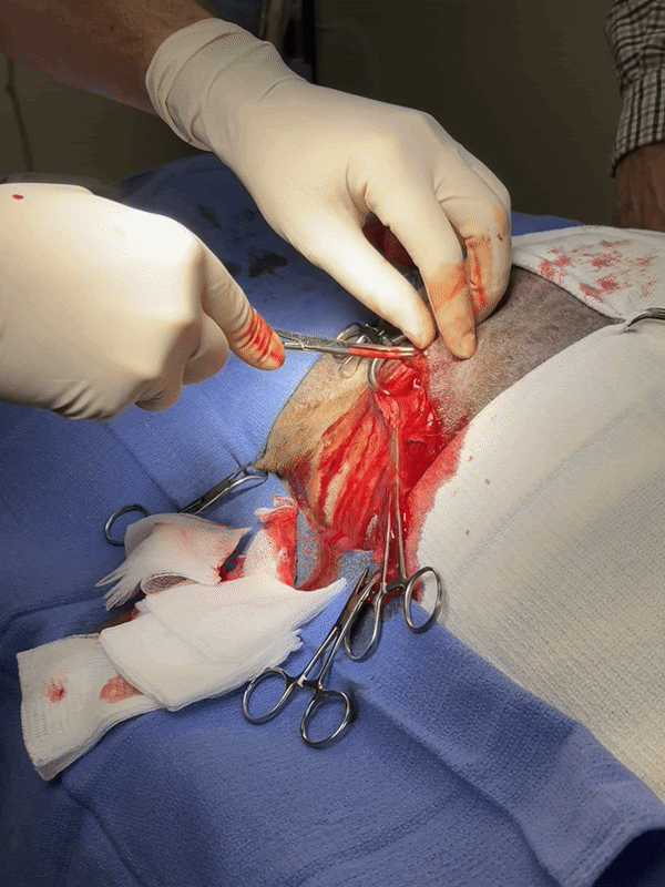
Outcome:
Surgery was performed to remove the foreign body via the abscess over the right caudal ribs. A wooden linear object (large toothpick or similar) was removed. An exploratory laparotomy was not immediately performed. Though there were no visible wounds, external penetration was suspected due to the orientation of the pointed part of the foreign object being deeper into the subcutaneous tissues. The patient made a full recovery following surgery.

Discussion:
Ultrasound can be used to routinely assess internal abdominal structures, but can also assess more superficial soft tissue structures such as abnormal subcutaneous and muscle tissues. While differentials for this patient initially included other causes of fever of unknown origin that may be commonly identified via abdominal ultrasound, an extra-abdominal abnormality was identified using the same imaging modality in an unexpected location.
Special thanks to the staff at Varina Veterinary Clinic for collaborating on this case.