
August's Case of the Month- 2022
Visceral Mast Cell Tumor
PATIENT INFORMATION:
Age: 11yo
Gender: Female Spayed
Species: Canine
Breed: Golden Retriever
Weight: 63lbs
HISTORY:
Previously healthy. Initially presented for vomiting and diarrhea with a normal appetite. Treated for clostridium and spirochetes with supportive care, probiotics and metronidazole. Barium study performed with no evidence of obstruction or foreign material was also performed. Presented the next day to the emergency clinic for persistent vomiting and diarrhea. Repeat radiographs show reduced cranial abdominal detail and hepatomegaly. No free fluid noted on AFAST. Represented to primary veterinarian one week later with blood in stool, febrile with mild neutrophilia and monocytosis. No reported history of mass removals, FNAs or biopsies.
ULTRASOUND FINDINGS:
Liver - Moderate-severely increased size, rounded shape and moderately coarse hyperechoic echogenicity with an ill-defined hypoechoic small mass in the left liver. (3.1x2.7cm)
Spleen- The spleen was moderately enlarged(3.1cm), elongated, and rounded in shape with a coarse echotexture. There was a cavitated capsule altering small mass in the tail of the spleen(2.3x2.1cm)
Lymph nodes - Multiple mesenteric lymph nodes were moderate-severely enlarged with rounded shape having a mottled mildly hypoechoic echogenicity.(at least 7.6cm in length and 2.9cm in depth) The medial iliac lymph nodes were mildly plump with an isoechoic echogenicity. Lt/Rt: 0.8/0.7cm
Gastrointestinal Tract - The stomach had increased wall thickness with some areas of hypoechoic hazy loss of layering detail.(6.5-7.3mm) The pylorus was free of obstruction. Many loops of the small intestine had increased wall thickness with disproportionately large mucosal layers. Some loops had hazy hypoechoic loss of layering detail. There was echogenic stippling throughout the mucosal layers. The bowel had mildly increased overall motility with liquid contents. The colon had normal to mildly increased wall thickness and normal layering. No focal intestinal masses or obstructions identified. Duodenum 5.6-6.0mm, Jejunum 3.7-5.9mm, Colon 3.0mm
Mild ascites noted throughout the abdomen.
SELECT IMAGES:
Image 1: Splenomegaly and cavitated splenic lesion.
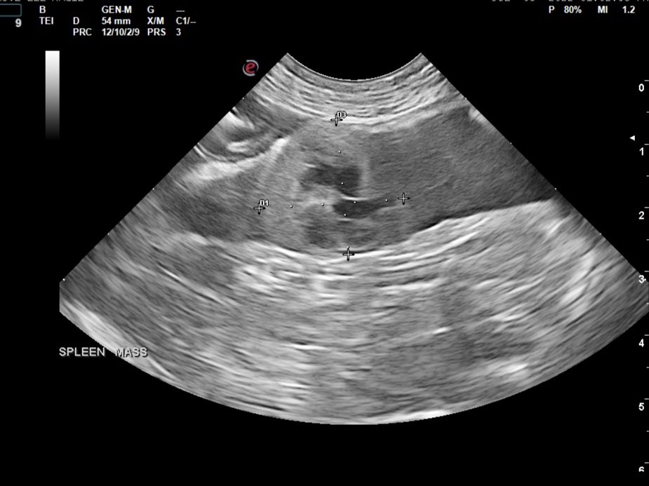
Image 2: Gastric wall thickening with hazy layering detail loss

Image 3: Severely enlarged and hypoechoic mesenteric lymph node.

Diagnostic tests performed:
Ultrasound guided fine needle biopsies of the spleen and mesenteric lymph nodes were collected.
Diagnosis
Spleen: Mast cell tumor
Mesenteric lymph node: Mast cell tumor
Cytology Comments
The diagnosis of a mast cell tumor is definitive (100% confidence), for both locations, consistent with a metastatic process. The slides from the two locations appear fairly similar, are moderately to highly cellular and consist of few to many red blood cells and a nucleated cell population predominated by mast cells with few small lymphocytes and rare plasma cells. The mast cells have a moderately to well granulated cytoplasm that surrounds a centrally located round nucleus with loosely clumped chromatin pattern. Anisokaryosis and anisocytosis are mild to moderate. Many purple granules are present in the background.
Cytology Images:

Image 4: Lymph Node aspirate - Several mast cells with fewer small lymphocytes and a plasma cell*Image provided by Eastern VetPath

Image 5: Speen- Numerous mast cells*Image provided by Eastern VetPath
CASE OUTCOME:
Patient was started on steroids however her clinical signs progressed and due to her grave prognosis she was humanely euthanized the following day.
PROGNOSIS/DISCUSSION:
The diagnosis of primary visceral mastocytosis is uncommon in canine patients. The liver, spleen and gastrointestinal tract are the most common sites for primary visceral MCT. It is possible that the splenic mass in this case was the primary tumor. The prognosis in these cases is poor and diagnosis is often delayed due to non-specific clinical signs(Vomiting/Diarrhea/Anorexia)
References:
Garrett L. Canine mast cell tumors: diagnosis, treatment, and prognosis. Vet Med (Auckl). 2014;5:49-58
https://doi.org/10.2147/VMRR.S41005
Takahashi T, Kadosawa T, Nagase M, Matsunaga S, Mochizuki M, Nishimura R, Sasaki N. Visceral mast cell tumors in dogs: 10 cases (1982-1997). J Am Vet Med Assoc. 2000 Jan 15;216(2):222-6. doi: 10.2460/javma.2000.216.222. PMID: 10649758
Special thanks to Dr. Emily Fant at Shady Grove Animal Clinic and Dr. Casey Leblanc at Eastern VetPath.